
 A CT scan (or CAT scan) is a form of X-ray scan which uses a process called computerized tomography to build up detailed images of the interior of a person’s body. Such a scan can be used to look at the internal organs or bones in order to check for major structural health issues. The result is a sequence of images slices which show cross-sections of the body. These are normally examined by an expert.
CT scans use X-rays, and because the internal tissues do not show up clearly on X-ray photographs, it is commonly necessary to inject a substance called a contrast agent into the patient. This is often a chemical based on iodine which makes the internal tissues more opaque to X-rays. In addition, if the patient is being scanned because of suspected intestinal problems, they will be asked to drink quite a large quantity of liquid contrast agent before having the scan. This fills the gut with material that is opaque on the CT scan, making the shape of the intestines more clearly visible.
Exposure to X-rays carries an increased risk of cancer. In addition, injected contrast agent can sometimes cause allergic reactions. For some people who are known to be allergic to the contrast agent, it is necessary to suppress the immune system by taking prednisone pills for a couple of days prior to the scan and also taking antihistamines.
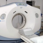
The experience of having a CT scan involves lying on a flat bed which can be slid inside a large donut-shaped machine. The machine contains rotating parts which make quite a lot of noise. Normally it takes only about 20 minutes to complete a CT scan. During this time the patient may need to receive an injection of contrast agent, so this involves also placing an IV into a vein. A remote controlled machine injects the contrast dye at the time of the scan.
CT scans can show intestinal dilation due to obstruction and may show narrowing due to Chron’s disease. They can detect the presence of foreign objects, indicate the shape and position of your colon, and also are used to image the appendix to make sure it is not inflamed.