
 In order to understand what can go wrong with digestion it is important to have an appreciation of the normal process by which food is processed by the body.
To stay alive, humans need three things: an energy source, which is derived from sugars, complex sugars, or fats; amino acid building blocks for growth, which come from the breakdown of proteins; and various essential vitamins and minerals. The digestive system provides these nutrients from the many sources of food that are available.
The first step of food consumption involves the mouth. Food is chewed by the teeth which breaks it into small pieces, and this is important because it increases the surface area, allowing more rapid digestive processes. Salivary glands produce saliva which contains the digestive enzymes amylase and lipase. Amylase starts the process of breaking down starch into simple sugars, and lipase acts to decompose fatty triglycerides into glycerides and fatty acids. The saliva wets the food and allows the tongue to shape it into a “bolus” which is easily swallowed. Saliva keeps the surface of the mouth healthy and moist and also contains various antimicrobials which prevent the overgrowth of oral bacteria.
After it has been swallowed the food moves down the esophagus which is the first part of the 30 foot long tube of the gastrointestinal tract that connects mouth to anus. The whole gut is a tube made of smooth muscle. This muscle can contract under the control of the nervous system in such a way as to create waves of peristaltic motion that propel food further down. The nerves can sense the pressure from a lump of food and produce a wave of contraction behind the lump and a wave of relaxation ahead of it, in such a way that it moves the food forwards. Gravity is not required, and so astronauts can still digest food without any problem while floating in space. These peristaltic waves move the food down the esophagus into the stomach.
The primary wave of contraction in the esophagus after swallowing propels the food forward but it moves at a fixed rate, and may overtake or lag behind that actual food position. If the nerves of the gut sense that the food is still present, they will continue to generate further waves of motion to try to move it onwards. In addition, when no food is present, spontaneous waves of activity are generated, called the migrating motor complex. These act to repeatedly clean out the gut and remove smaller particles of food and excess bacteria, and are a sort of housekeeping function.
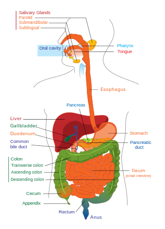 The esophagus empties food into the top of the stomach through the lower esophageal sphincter (cardiac sphincter) which is a ring of muscle that automatically opens to allow food to pass. Its purpose is to prevent any flow of stomach acid (acid reflux) back up into the esophagus, for example while lying down, which might lead to damage to the gut lining, inflammation, and the pain of heartburn.
The stomach is a large muscular container which can hold about 1 liter of food. Glands in the walls of the stomach secrete gastric juices in the form of hydrochloric acid and digestive enzymes which provide an environment conducive to the breaking down of food into constituent parts which are more easily digested. The secreted enzymes include proteases which break proteins down into peptides and amino acids. The strongly acid environment of the stomach is important because it kills off most microorganisms and is also needed for the proper activation of the protease enzymes. Mucous is secreted by the stomach wall and provides a protective layer against the corrosive effects of gastric acid. The muscles of the stomach contract repeatedly in order to churn the contents and ensure that they are thoroughly blended. The final result is a partially digested mixture known as chyme. The stomach may hold onto the food from 40 minutes to a couple of hours depending on what has been consumed, but eventually releases the chyme slowly through the muscular valve of the pyloric sphincter into the duodenum of the small intestine where it begins to be absorbed.
The majority of digestion takes place in the small intestine and involves the breakdown of various food substances into water soluble forms that can easily be carried away by blood plasma. The main function of this part of the gut is to provide a protective membrane with a large surface area in contact with digested food through which nutrients can be readily absorbed.
After leaving the stomach, the chyme mixture passes into the duodenum which is the first part of the small intestine. Here it mixes with digestive enzymes that are excreted by the pancreas and with bile that is excreted by the liver. The digestive enzymes are chemicals that facilitate the breaking down of complex sugars (such as carbohydrates) into simple sugars, proteins and peptides into amino acids, and fats into fatty acids. Bile is a surfactant that helps to emulsify fatty substances so that they break up into many tiny globules which can more easily interact with the gut wall. Some of the bile that is generated by the liver is also stored in the gall bladder. This organ contracts to release bile when fatty food is present and adds to other bile which is continuously entering the duodenum from the liver. In the duodenum, specialized glands, known as Brunner’s glands, secrete mucous and also bicarbonate in order to neutralize stomach acids. In addition, various digestive enzymes and protective mucous are a normal constituent of secretions throughout the small intestine.
Food progressively moves from the duodenum into the jejunum and ileum where it continues to be digested and absorbed via the intestinal surface. In order to facilitate this process the gut wall is structured so that it has many small protrusions known as villi. These bumps are easily seen if you examine tripe – a food made from cow intestine that is sometimes eaten in Vietnamese soups. In addition, each epithelial cell that makes up the intestinal wall has smaller microvilli in its cellular membrane that form a surface called the brush border. The villi and microvilli drastically increase the surface area for absorption. Nutrients absorbed in the intestine are transported by blood circulation to the liver for further processing and storage.
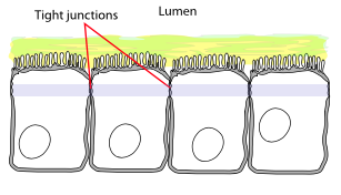 The cells that constitute the intestinal wall form a protective barrier which strategically controls which chemicals can cross from the interior of the gut (lumen) into the blood stream. In particular, large molecules, proteins, or intestinal bacteria and their metabolites, are prevented from passing, whereas the healthy products of digestion are transported across. An important part of this structure are the tight junctions which are the contact boundaries between intestinal epithelial cells. These are normally virtually impenetrable to fluids, although their permeability has recently found to be modulated by a protein called zonulin.
The final section of the small intestine is the ileum. An important function of the ileum is the absorption of vitamin B12 and bile salts. The bile salts are transported back to the liver to be secreted again (bile circulation). The final part of the ileum (the terminal ileum) connects to the first part of the large intestine (the cecum of the colon) via the ileocecal valve. This valve opens to let food exit the small intestine and helps prevent the contents of the cecum, which consists of large numbers of bacteria, from refluxing back into the small intestine and interfering with digestion.
The main function of the large intestine (colon) is to store waste and to absorb water and electrolytes from the flow of material exiting the ileum. It also provides a place for bacterial fermentation by symbiotic bacteria which, by feeding on indigestible matter, produce additional nutrients that can be used by the body. The colon consists of the cecum, ascending colon, transverse colon, descending and sigmoid colon, and the rectum. The appendix is a finger-like protrusion attached to the bottom of the cecum. It has traditionally been viewed as a non-functioning (vestigial) organ, even though it does contain important immune-related lymphoid tissue. Recently it has been proposed that the appendix maintains a sample of the normal gut bacteria which is important for re-innoculating the colon with healthy bacteria following a food poisoning event.
The colon is also a dumping ground for any substances that the body wishes to get rid of which are not excreted in the urine.
Material transported through the colon eventually finds its way to the rectum which acts as a temporary storage site for feces. As the rectum becomes full, stretch receptors in the walls send nerve signals which produce the urge to defecate. Defecation involves relaxing the internal and external sphincter muscles and the induction of peristaltic motions in the rectum which propel material out through the anus.
The process of digestion normally takes between 24 and 72 hours and adults on average have one bowel movement per day.
The process of digestion is controlled by the enteric nervous system, a distinct part of the nervous system which is able to operate largely independently from the central nervous system and brain. Most of the enteric nervous system is found in neural layers which surround the walls of the intestine. Sensory neurons in these layers are able to monitor nutritional factors and the pressure from food within the intestine. Motor neurons control the contraction of muscles in the gut wall and generate various forms of peristaltic waves that mix or propel food along the intestinal tract. The majority of nerves connecting the gut to the brain are for relaying the sensations of digestion, but others modulate the digestive process. The enteric nervous system communicates with spinal nerves, which are associated with sympathetic (fight and flight) activity, and also with the vagus nerve, which is associate with parasympathetic (rest and digest) activity.
Bacteria play an important role in digestion and different regions of the gut have different bacterial profiles. In the mouth, the dominant species are streptococci, lactobacilli, staphylococci, corynebacteria, and various anaerobes such as bacteroides. The oral bacterial population is controlled by antibacterial secretions in the saliva. If the right bacteria are present, this helps prevent colonization by pathogens such as yeasts.
 The stomach is a hostile place for bacteria and so only a few acid-tolerant species survive the passage through this region. The small intestine is largely sterile because excess bacteria would compete for nutrients at the digestive surfaces. Lactic acid bacteria are more common in this area and help to create an acid environment which is less conducive to the proliferation of pathogens. The migrating motor complex which regularly sweeps a propulsive wave through the small intestine is part of the body’s system for clearing out any excess bacterial growths following the digestion of a meal. The number of bacteria per milliliter of fluid increases towards the end of the ileum and becomes very large as one enters the colon. Many hundreds of species of bacteria have been identified in the colon. These predominantly include anaerobic bacteria, and four types dominate: Firmicutes, Bacteroidetes, Actinobacteria, and Proteobacteria. Some of these bacteria perform useful functions which benefit the body. For example butyrate is an important chemical which is produced during bacterial fermentation of dietary fiber, and is a fuel which is used by the wall of the colon, and also modulates inflammatory processes.
A complex symbiosis exists between the body and the microflora of the gut contents. The waste within the colon contains enough bacterial toxins to kill the host many times over. Therefore layers of defense must be present at the intestinal wall to maintain appropriate separation between the sterile blood stream and the bacteria-filled lumen. A major defense is the mucous layer. Mucous is constantly excreted to lubricate the colon and to mechanically prevent bacteria from invading the body. In addition, immune factors such as secretory IgA provide an important antimicrobial activity, selecting for those beneficial bacteria which are minimally pathogenic to humans.
In summary, the intestinal tract is a highly balanced system which has evolved to efficiently extract the maximum nutritional value from food while ensuring health and protection for the individual.